Advanced Retina Care
Retina
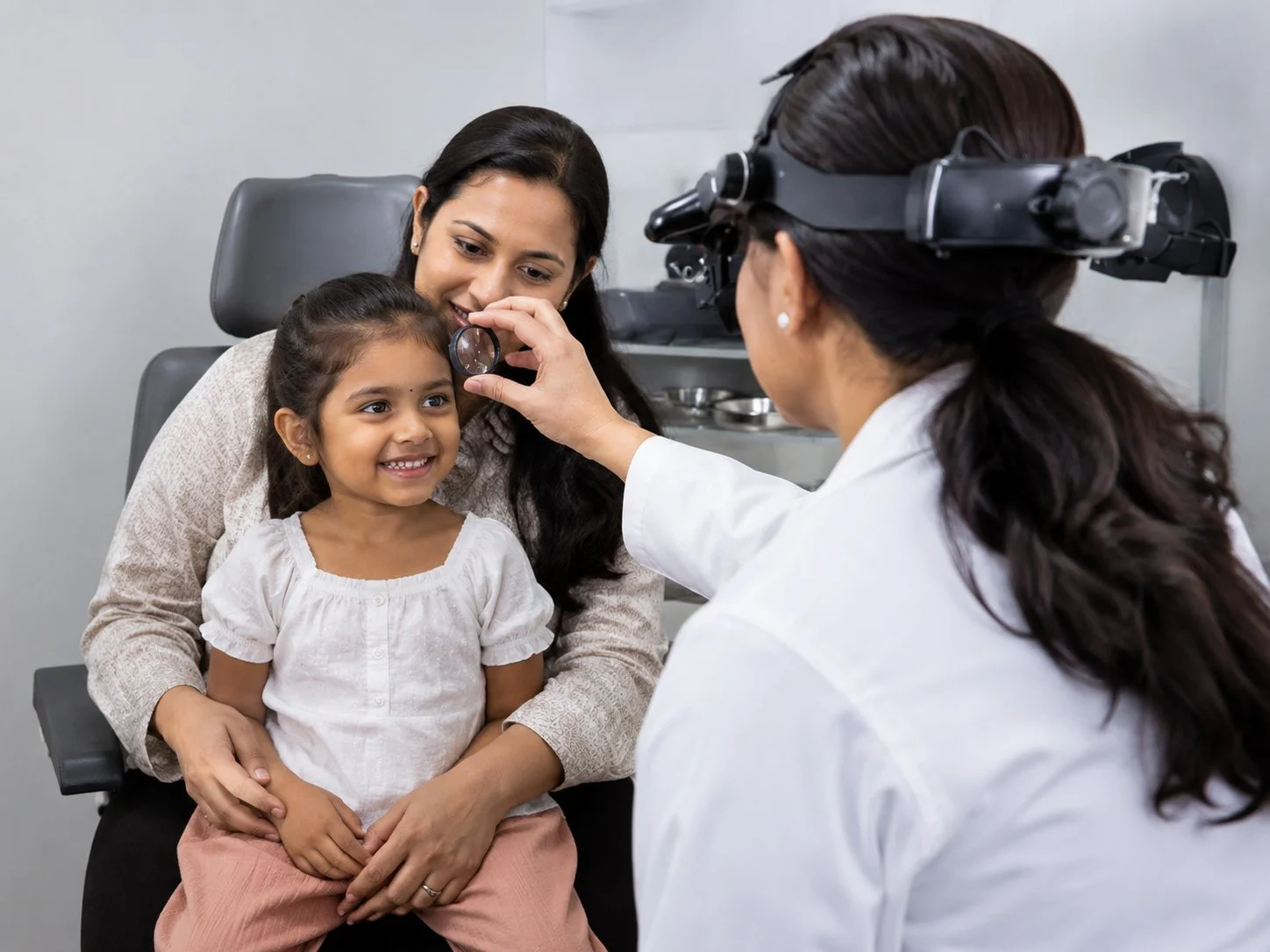
At NAMAH, retina care is built on detailed clinical examination, imaging-based diagnosis, careful counselling and stage-wise treatment planning. We understand that retinal disorders often need precision, patience and close monitoring — where timely follow-up can be just as important as the first consultation.
What is the Retina?
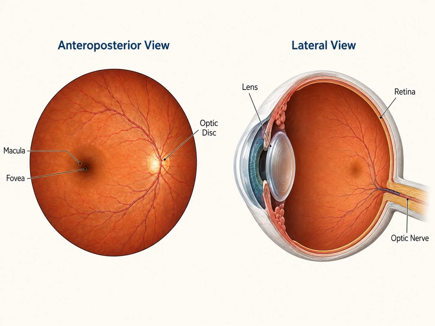
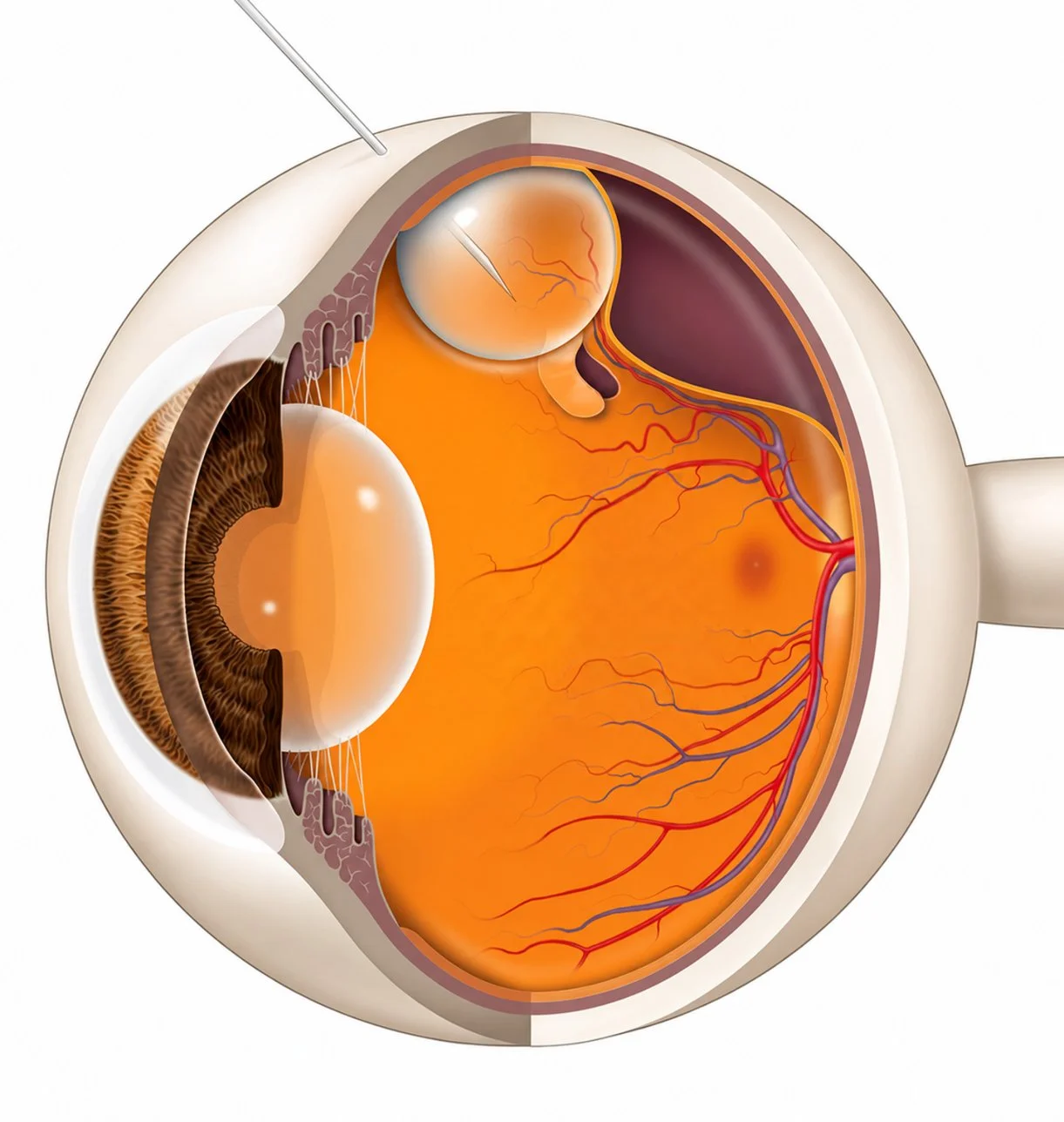
The retina is the delicate nerve layer at the back of the eye. It receives light and sends visual signals to the brain via the optic nerve.
Retinal diseases can result in blurred, distorted, patchy or sudden decrease in vision
Some retinal diseases are silent in the early stages. Others need urgent treatment to prevent permanent damage.
That is why a retina evaluation is not just a routine eye check - it is a detailed examination of the most sensitive part of the eye that actually captures vision.
When should you see a retina specialist ?
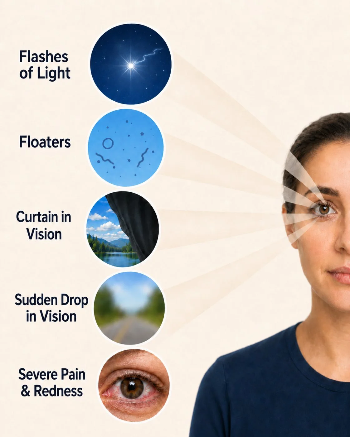
Emergency evaluation in case of sudden increase in floaters or black spots, flashes of light, a curtain/shadow in vision, sudden drop in vision, severe eye pain and redness.
Patients with gradual decrease in vision, distorted lines, dark central patch
Diabetic patients for Diabetic retinopathy screening
Elderly patients (ideally over 50 years of age) for Age related macular degeneration screening
High myopes for baseline and follow up screening for peripheral retinal degenerations
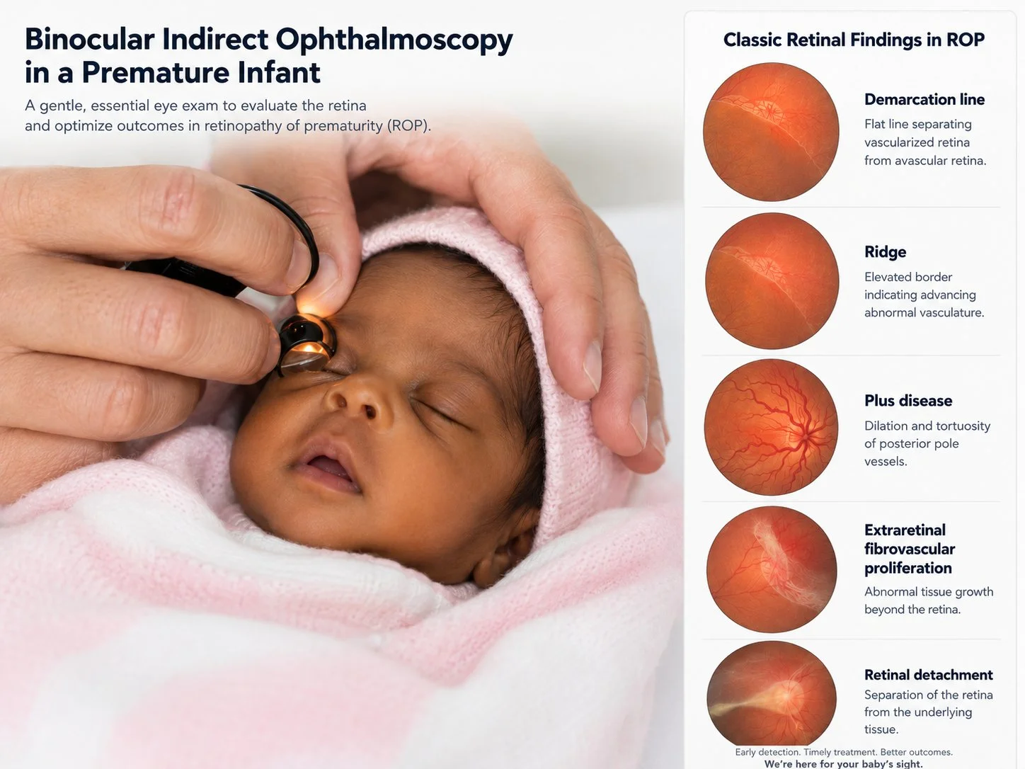
Premature baby needing ROP screening
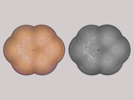
Click on the image to zoom in
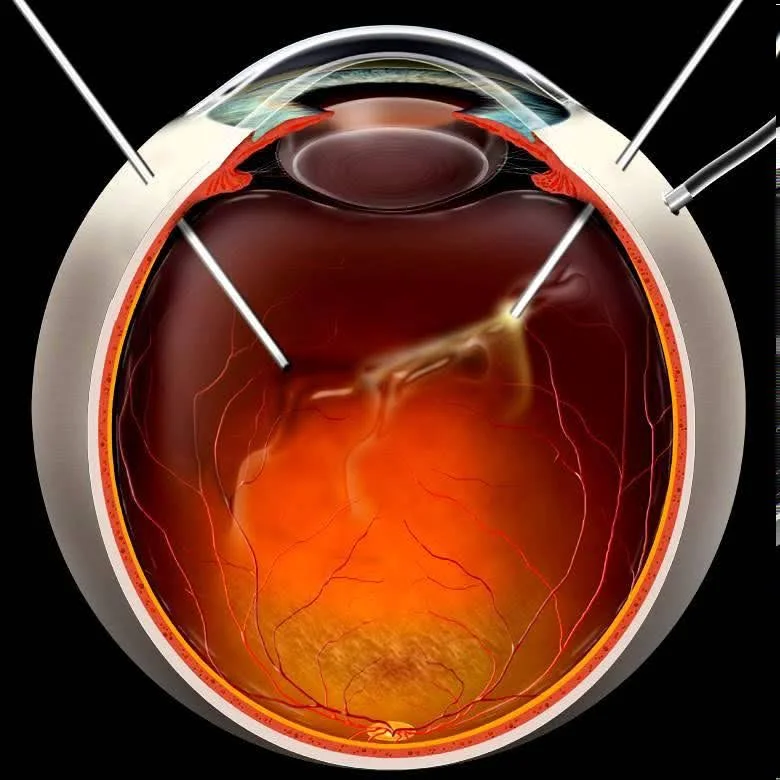
What does a Retina specialist at Namah evaluate ?
From symptom to treatment plan - a clear retina care pathway.
Symptoms, ocular and medical history along with systemic history of the patient are noted
Symptoms and history
Vision testing
Anterior segment examination
Dilating eye drops instilled (20-30 mins waiting period)
Dilated retina examination
Imaging if required
Management options and care explained
Documentation and follow up schedule explained to patient.
At Namah, every patient undergoes a comprehensive evaluation right from vision assessment to anterior and posterior segment evaluation. Retina care especially relies on detail. A patient undergoes a detailed retinal examination after dilatation. Imaging modalities are further used to understand the disease depth, activity and progression better.
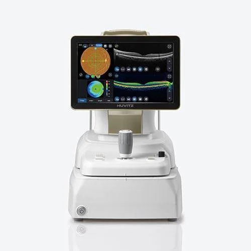
Namah is well-equipped with advanced diagnostics for retinal imaging
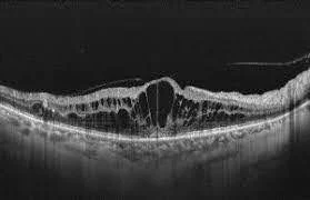
Huvitz OCTavius SD-OCT(Spectral domain-Optical coherence tomography)
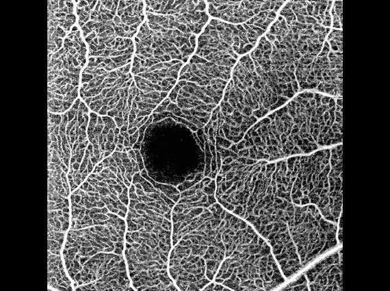
Spectral domain-Optical coherence tomography uses light for speedy and detailed retinal layer imaging allowing for detection of subtle changes as well as for objectively monitoring disease progression and treatment efficacy.
OCTA (Optical coherence tomography- Angiography )
Optical coherence tomography- Angiography technology allows for non-invasive (without the use of a dye injection) visualisation of the blood vessels of the eye and any disease affecting them. This is extremely useful to differentiate between normal blood vessels and abnormally formed new vessels in the eye which can lead to bleeding and scarring in the eye in advanced cases.
Many retinal conditions can be managed with targeted treatment when detected at the right time.
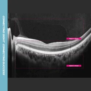
Click on the image to zoom in
Common Retinal Conditions
Diabetic Retinopathy and Diabetic Macular Edema
Diabetes can damage the small blood vessels of the retina. Diabetic retinopathy is one of the most important reasons for regular retinal screening. In early stages, a person may see well and still have retinal changes. As the disease progresses, it can cause bleeding, swelling in the central retina, new abnormal blood vessels, vitreous hemorrhage or traction on the retina. Timely diagnosis thus, becomes very important in order to prevent severe complications.
At NAMAH, diabetic retina care focuses on early detection, OCT-based evaluation, retinal imaging, stage-wise treatment planning and follow-up documentation. Depending on the stage, treatment may include observation, better systemic control, retinal laser, intravitreal injections or surgery.
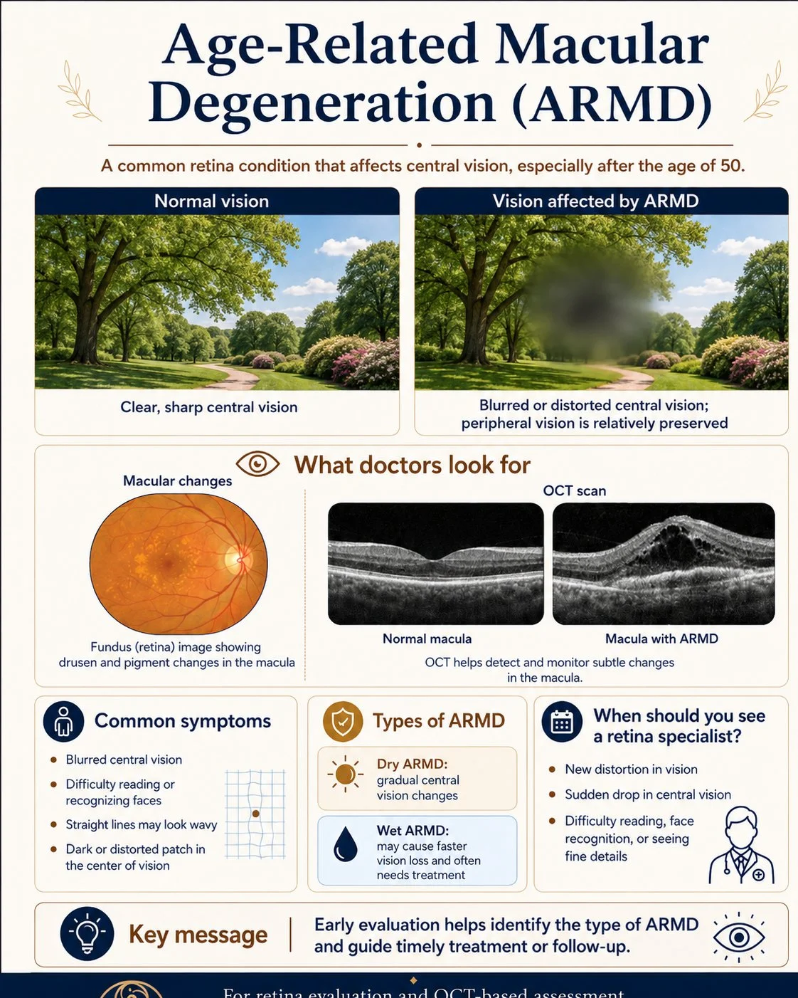
Age-Related Macular Degeneration(ARMD)
The macula is the central, most sensitive part of the retina responsible for reading, recognizing faces and fine detail. Age-Related Macular Degeneration affects central vision, reading and face recognition in elderly individuals. Clinical and OCT evaluation helps to identify dry or wet forms which requires treatment in the form of intravitreal injections.
Retinal Vein Occlusion
A blockage in retinal veins can cause sudden blurred vision or bleeding/swelling in the retina. More common in individuals with hypertension.
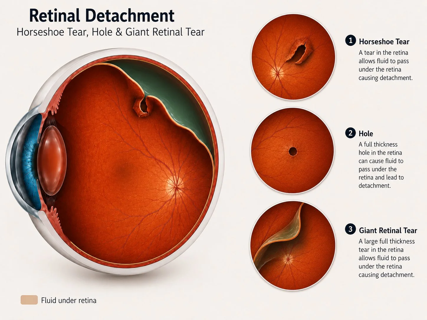
Retinal Tears and Holes
Small breaks in the retina may need timely laser to reduce the risk of retinal detachment.
Retinal Detachment
A serious condition where the retina separates from its support layer. Sudden floaters, flashes or a curtain-like shadow need urgent attention.
Vitreous Hemorrhage
Bleeding inside the eye can cause sudden hazy vision or black floaters and often needs detailed retinal evaluation.
Macular Hole
A defect in the central retina that can cause distortion or central vision loss. Some cases require surgery.
Myopic Retinal Degeneration
High myopia can stretch the retina and increase risk of peripheral retinal weakness or macular changes including a macular hole.
Inherited Retinal Conditions
Evaluation, documentation, genetic counselling and referral support is provided in patients with genetic eye diseases.
ROP Screening
Retinal examination for premature infants with timely referral for appropriate treatment to prevent vision threatening complications including blindness.
Pediatric Retinal Evaluation
Assessment for abnormal retinal reflex, poor visual response, prematurity-related retinal disease and selected pediatric vitreoretinal concerns.
Click on the image to zoom in
Treatment modalities at Namah
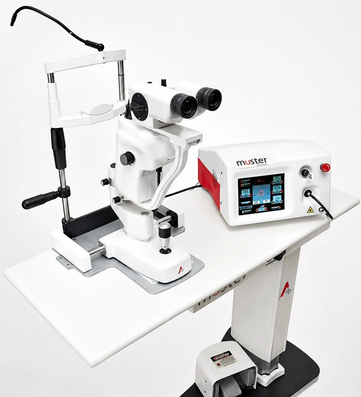
Retinal Laser
Focused LASER light treatment applied to the retina as an OPD procedure under topical anesthesia (using eye drops)
When: Retinal tears, diabetic retinopathy, proliferative vascular conditions and ROP.
At Namah, multispot green laser (AEON meditech) is used to deliver multiple laser spots at one time, ensuring quick laser procedure completion with minimal pain for the patient.
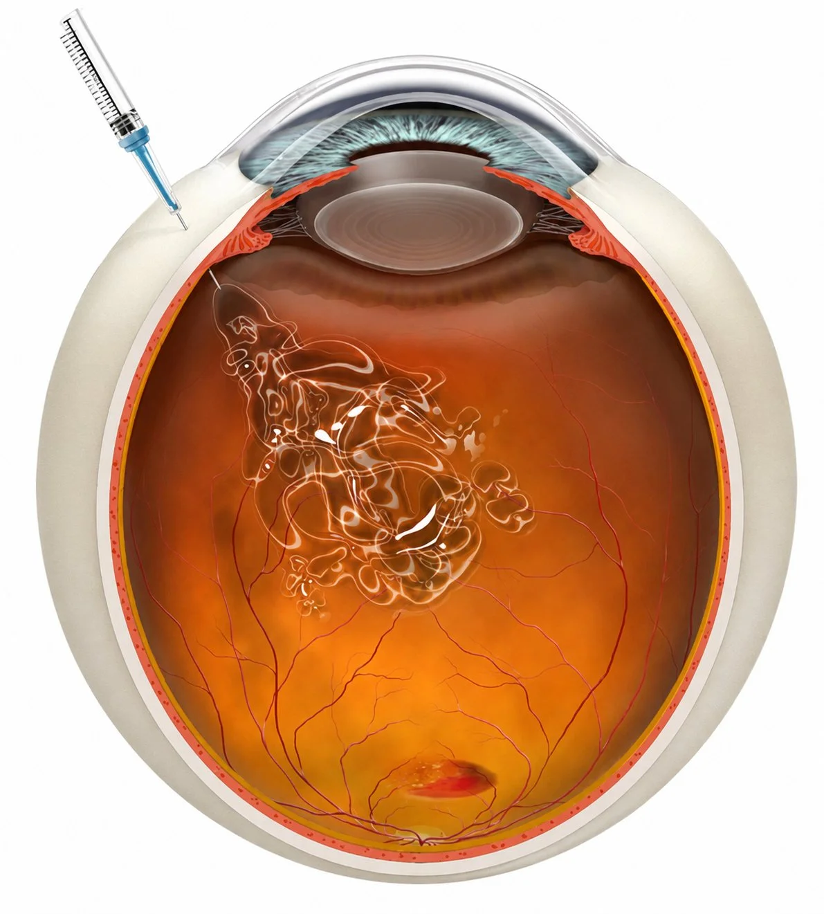
Intravitreal Injections
Medicine delivered inside the eye for retinal swelling or due to presence of abnormal new blood vessels. This is performed with sterile precautions under topical anesthesia in the modular operation theatre.
When: diabetic macular edema, wet AMD, vein occlusion-related swelling, proliferative vascular diseases and selected inflammatory conditions.
Vitrectomy Surgery
Advanced Minimally Invasive Vitrectomy Surgery (MIVS) is performed for vitreoretinal problems such as Retinal detachment, non-clearing vitreous hemorrhage, macular hole and diabetes complications, offering precise and usually sutureless surgery for complex conditions while ensuring optimal results and faster patient recovery. In some cases, silicone oil or gas is placed inside the eye during surgery to support the retina. Silicone oil removal may then be required after a few months. Need for special post- operative care in the form of head positioning may be needed in certain cases
At Namah, the Oertli OS 4 vitrectomy platform is used for vitrectomy surgery. It is an advanced surgical platform with precise and controlled surgical environment that allows for stable ocular dynamics ensuring improved surgical safety. -MIVS incisions are very small and usually do not require any sutures. -Faster healing and better visual outcomes.
Scleral buckling
Scleral buckling is done in selected cases for retinal reattachment and support. In this procedure, a soft silicone band is placed around the eye.
Pneumatic retinopexy (PNR)
PNR is a safe, quick, minimally invasive procedure involving the injection of a gas bubble followed by laser to attach certain types of retinal detachments. The patient is then advised appropriate head positioning at home. In cases with successful retinal re-attachment, there may be no further need for a vitrectomy surgery.

Click on the image to zoom in
Frequently Asked Questions
I have diabetes but my vision is normal. Do I still need a retina check?
Yes. Diabetic retinopathy can begin before vision changes. A dilated retinal examination and imaging, when needed, can detect early disease and guide follow-up.
How often should diabetic patients get retina evaluation?
Most diabetic patients need at least yearly retinal evaluation, but the interval may be shorter if retinopathy, macular edema, pregnancy, kidney disease, uncontrolled sugar or previous treatment is present.
Are eye injections painful?
The eye is numbed before an intravitreal injection. Most patients feel pressure or mild discomfort rather than significant pain. Sterile precautions and aftercare instructions are important.
Are floaters dangerous?
Occasional long-standing floaters may be harmless, but a sudden increase in floaters, flashes or a shadow in vision can indicate a retinal tear or detachment and should be checked urgently.
What symptoms suggest retinal detachment?
Sudden floaters, flashes of light, a curtain-like shadow, side vision loss or sudden drop in vision are warning symptoms that need urgent retina evaluation.
What is ROP?
ROP is Retinopathy of Prematurity, a retinal condition seen in premature babies where retinal blood vessels may grow abnormally. Screening is important because parents may not notice symptoms early.
When should premature babies be screened for ROP?
ROP screening timing depends on birth weight, gestational age and neonatal risk factors. In India, babies born at or before 34 weeks, weighing 2000 grams or less, or clinically unstable babies are commonly considered for screening. The first screening should be scheduled as advised by the neonatologist/ROP specialist and should not be delayed.
Can uveitis come back?
Yes. Some types of uveitis can recur or become chronic. Follow-up is important even after symptoms improve.
Is uveitis related to other body diseases?
Sometimes. Uveitis may be associated with infections, autoimmune conditions or inflammatory disease elsewhere in the body. Selected patients may need blood tests or physician/rheumatology coordination.
What is OCT?
OCT is a non-invasive scan that gives cross-sectional images of the retinal layers. It helps detect swelling, fluid, macular disease and treatment response.
Is retinal laser painful?
Many patients tolerate retinal laser well with numbing drops. Some may feel mild pricking, brightness or discomfort depending on the area treated.
Can retina diseases be treated without surgery?
Many retina conditions can be managed with observation, medicines, laser or injections. Some conditions such as retinal detachment, macular hole, advanced diabetic traction or non-clearing bleeding may need surgery.
When should I seek emergency retina care?
Sudden flashes, many new floaters, curtain-like shadow, sudden vision loss or eye trauma should be treated as urgent.