
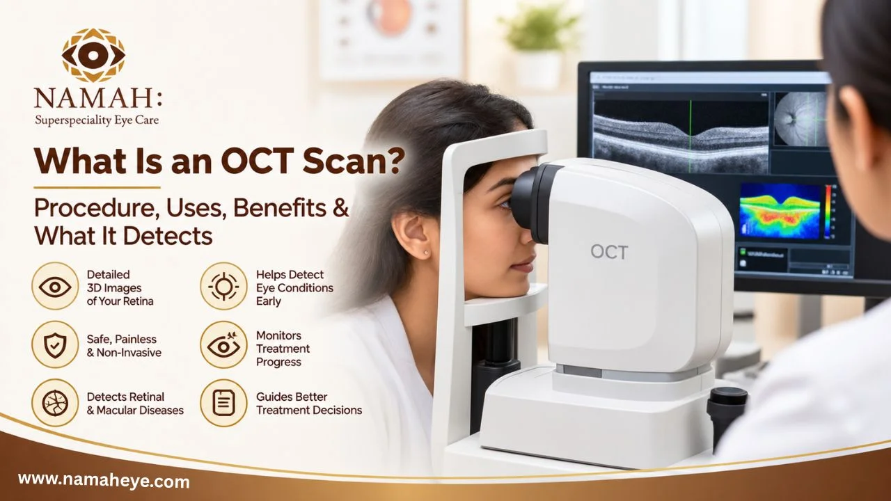
What Is an OCT Scan? Procedure, Uses, Benefits & What It Detects
Have you ever been told that you need an OCT scan for your eyes and wondered what it actually means?
OCT, or Optical Coherence Tomography, is a modern, non-invasive eye imaging test that allows an eye specialist to see detailed images of the different layers at the back of your eye. Unlike a regular eye examination, an OCT scan can provide a much closer look at the retina, macula and, in some cases, the optic nerve.
The test is quick, does not require the eye to be touched, and uses light rather than X-rays. It is commonly used to help doctors detect, diagnose and monitor several retinal and optic nerve conditions.
But what does an OCT scan actually show, and when do you need one? Let’s understand it in simple terms.
What Is an OCT Scan?
OCT stands for Optical Coherence Tomography.
An OCT scan is an imaging test that creates detailed cross-sectional pictures of the structures inside your eye. It uses reflected light to capture images of the retina layer by layer.
Think of it as taking a very detailed “section-by-section” picture of your eye.
A normal eye examination allows the doctor to look at the overall health of your eyes. An OCT scan provides additional information by showing the thickness and structure of individual retinal layers.
This can help an eye specialist identify changes that may not be easy to see during a routine examination.
Why Is an OCT Eye Scan Important?
The retina is located at the back of the eye and plays a major role in vision. The macula, which is part of the retina, is responsible for detailed central vision.
Small changes in these structures can affect your ability to read, recognize faces, see fine details or maintain clear vision.
An OCT scan can help doctors:
- Examine the retina in detail
- Identify swelling or fluid
- Check changes in retinal thickness
- Examine the macula
- Monitor retinal conditions over time
- Assess certain optic nerve changes
- Compare current images with previous scans
This makes OCT particularly useful when an eye specialist needs more information than a standard eye examination can provide.
What Can an OCT Scan Detect?
An OCT eye test can be useful in evaluating several eye conditions. It does not replace a complete eye examination, but it can provide important information that supports diagnosis and monitoring.
1. Diabetic Retinopathy
Diabetes can affect the small blood vessels of the retina.
An OCT scan can help identify retinal changes and detect swelling or fluid in the macula, which can occur with diabetic macular edema.
For people with diabetes-related eye problems, OCT may also be useful for monitoring changes over time.
2. Age-Related Macular Degeneration
Age-related macular degeneration, or AMD, affects the macula and can impact central vision.
An OCT scan can provide detailed images of the macula and help the doctor identify structural changes, including fluid or other abnormalities.
It can also help monitor the eye over time when ongoing treatment or observation is required.
3. Glaucoma
OCT can also be used to examine parts of the optic nerve and measure retinal nerve fibre layers.
In people being evaluated for glaucoma, OCT findings may be considered along with eye pressure measurement, optic nerve examination and visual field testing.
OCT alone does not diagnose glaucoma. It is one part of a complete assessment.
4. Macular Hole
A macular hole is an opening that develops in the central part of the retina.
Because OCT provides cross-sectional images of the macula, it can help the doctor identify and assess a macular hole.
5. Epiretinal Membrane
An epiretinal membrane is a thin layer of tissue that can form over the surface of the retina.
It may cause blurred or distorted central vision. OCT can help the specialist see the membrane and understand how it is affecting the retina.
6. Retinal Swelling
Fluid accumulation within or under the retina can affect vision.
OCT can help identify areas of retinal swelling and allow the doctor to monitor whether the condition is improving, remaining stable or changing over time.
7. Other Retinal Conditions
Depending on the patient’s symptoms and clinical examination, OCT may also assist in evaluating conditions such as retinal vascular problems, central serous chorioretinopathy, vitreomacular conditions and other macular disorders.
When Does a Doctor Recommend an OCT Scan?
An OCT scan is not necessarily required for every person during every eye examination.
Your eye specialist may recommend an OCT eye scan when:
- You have unexplained changes in vision
- You have diabetes
- You have symptoms suggesting a retinal or macular problem
- You are being evaluated for glaucoma
- You have an existing retinal condition
- You need monitoring during retinal treatment
- You have age-related macular concerns
- You have high myopia or other risk factors
- Your doctor notices something during a routine eye examination that needs closer evaluation
The decision depends on your symptoms, eye examination and individual risk factors.
How Is an OCT Scan Done?
One of the biggest advantages of an OCT scan is that the procedure is simple and generally comfortable.
During the test:
- You sit in front of the OCT machine.
- Your chin is placed on a support.
- You look at a small target or light.
- The machine scans your eye without touching it.
- Detailed images of the retina and other structures are captured.
- The same process may be performed for the other eye.
The actual scanning takes only a short time. You simply need to keep your eyes focused and remain as still as possible while the image is captured.
Is an OCT Scan Painful?
No. An OCT scan is generally painless and non-contact.
Nothing needs to touch the surface of your eye during the scan. The machine uses light to create the images.
In some cases, your doctor may use dilating eye drops as part of the overall eye examination. If your pupils are dilated, you may experience temporary light sensitivity or blurred vision afterward.
Does an OCT Scan Use Radiation?
An OCT scan uses light-based imaging, not X-rays.
This is one of the reasons OCT has become an important imaging tool in modern eye care.
What Happens After an OCT Scan?
The OCT machine produces detailed images that are reviewed by your eye specialist.
Your doctor may look at:
- Retinal thickness
- Individual retinal layers
- Macular structure
- Areas of swelling or fluid
- Changes in the optic nerve
- Differences between your two eyes
- Changes compared with previous scans
If you have had an OCT previously, comparing older and newer scans can be particularly useful. It allows the doctor to monitor whether a condition is stable, improving or progressing.
Can You Understand Your OCT Report Yourself?
You may see different measurements, graphs, images and colour-coded areas on an OCT report.
However, an OCT report should not be interpreted on its own.
A result that appears abnormal on the report does not automatically mean that you have a serious eye disease. Similarly, an apparently normal image does not replace a complete eye examination.
Your eye specialist considers your OCT findings together with:
- Your symptoms
- Medical history
- Vision test
- Eye examination
- Eye pressure
- Previous scans
- Other diagnostic tests
This complete picture is important for an accurate diagnosis and treatment plan.
OCT Scan vs Regular Eye Examination
An OCT scan and a routine eye examination serve different purposes.
A regular eye examination allows the doctor to assess your overall eye health and vision.
An OCT scan provides detailed cross-sectional images of specific structures, particularly the retina and macula.
Therefore, OCT is best understood as an additional diagnostic tool rather than a replacement for a comprehensive eye examination.
In many cases, your doctor may recommend OCT because they need a more detailed view of a particular area of the eye.
What Are the Benefits of an OCT Scan?
The major benefits of an OCT eye scan include:
- Non-invasive: No instrument needs to touch your eye.
- Painless: The scan itself generally causes no discomfort.
- Quick: Images can be captured within minutes.
- Detailed: It provides layer-by-layer views of retinal structures.
- Useful for monitoring: Previous and current scans can be compared.
- Supports diagnosis: It provides additional information about retinal and optic nerve conditions.
- No X-ray radiation: OCT uses light-based imaging.
Is OCT Suitable for Everyone?
OCT is a widely used eye imaging technology, but whether you need one depends on your individual eye health.
A healthy person without symptoms or specific risk factors may not need an OCT scan at every visit.
On the other hand, people with diabetes, glaucoma-related concerns, retinal conditions, macular problems, high myopia or unexplained vision changes may benefit from OCT when recommended by their eye specialist.
Your doctor can determine whether an OCT scan is appropriate for you.
When Should You Get an Eye Check-Up?
Do not wait for a routine appointment if you experience sudden or unusual changes in your vision.
Contact an eye specialist if you notice:
- Sudden blurred vision
- Sudden reduction in vision
- New or increased floaters
- Flashes of light
- Distorted or wavy vision
- A dark spot or shadow in your vision
- Difficulty seeing central details
Some of these symptoms can be associated with retinal conditions that require prompt evaluation.
Frequently Asked Questions About OCT Scan
Is an OCT scan painful?
No. An OCT scan is generally painless and does not require contact with the eye.
How long does an OCT scan take?
The scanning process is usually completed within a few minutes, although the total eye-care visit may take longer if other examinations or pupil dilation are required.
What is an OCT scan used for?
An OCT scan is used to examine detailed layers of the retina and optic nerve and can help doctors diagnose or monitor conditions such as diabetic eye disease, macular disorders and glaucoma-related changes.
Is OCT the same as an eye test?
No. OCT is an imaging test. It is usually used alongside a comprehensive eye examination when your doctor believes detailed retinal or optic nerve imaging is needed.
Do I need an OCT scan every year?
Not necessarily. The frequency depends on your eye condition, risk factors, symptoms and your doctor’s recommendation. Some patients may need repeated scans for monitoring, while others may only need OCT when clinically indicated.
Conclusion
An OCT scan is a simple, non-invasive and detailed imaging test that helps eye specialists look beneath the surface and examine the different layers of the retina and other important eye structures.
It can play an important role in detecting and monitoring several retinal, macular and optic nerve conditions, particularly when combined with a complete eye examination.
If you have diabetes, glaucoma concerns, a known retinal condition, unexplained vision changes, or symptoms such as flashes, floaters or distorted vision, speak with an eye specialist to understand whether an OCT scan is appropriate for you.
For specialized eye evaluation and retinal care, you can consult Namah Superspeciality Eye Care for appropriate assessment and guidance based on your individual eye-health needs.