Advanced Eye Care

Uveal disorders

Uveal disorders of the eye include Inflammation of the Uveal tissue, which is one of the parts of the eye. It may be associated with infections, autoimmune conditions or inflammatory disease in the eye or elsewhere in the body.

What is the Uvea?

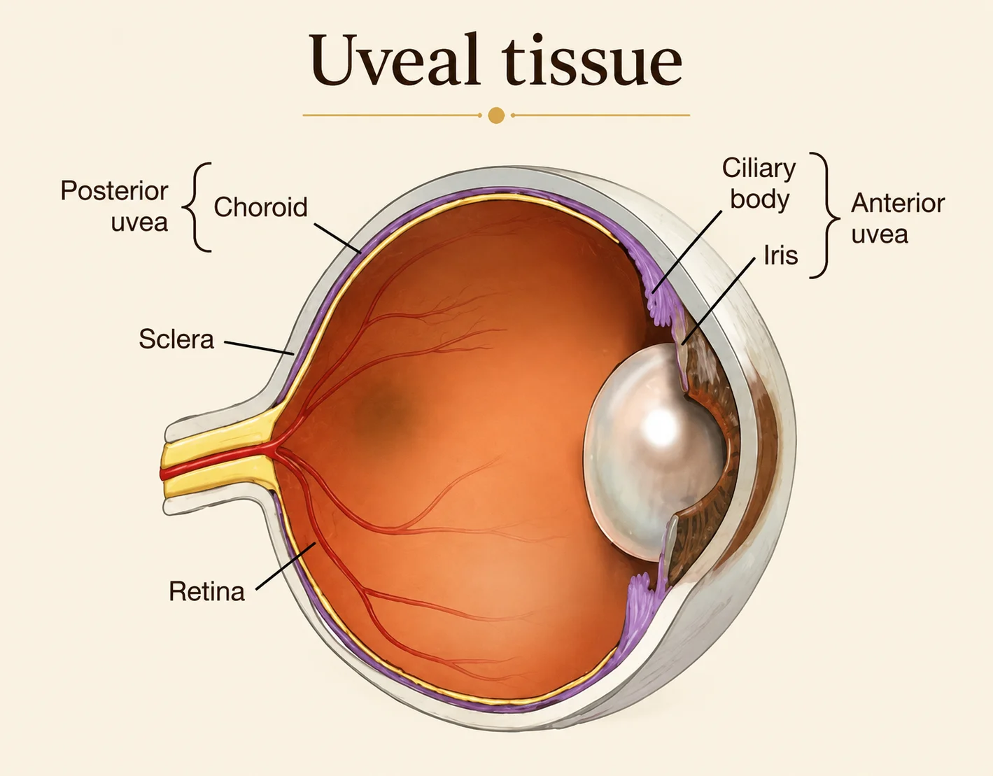
The uveal tissue is the pigmented, middle layer of the eye. It is located between the inner retina and the outer sclera. It consists of three main structures:

Iris

Colored part of the eye

Ciliary body

Muscles that are attached to the lens and alter the lens shape

Choroid

posterior part of the uvea containing a dense network of blood vessels

Common symptoms of Uveal disorders

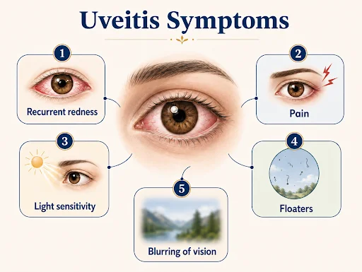
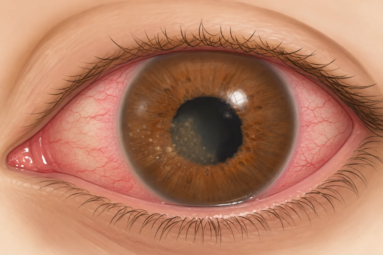
Patients may experience any of the following symptoms :

Redness

Eye pain

Blurring of vision

Light sensitivity

Floaters

Recurrent redness and pain

Click on the image to zoom in

Types of Uveitis

01
Anterior uveitis

Affects the anterior uveal tissue (Iritis) causing inflammation in the anterior chamber of the eye. This is commonly associated with pain, redness and light sensitivity

02
Intermediate uveitis

Affects the middle uveal tissue (ciliary body) and vitreous gel. This is associated with floaters, blurring of vision.

03
Posterior uveitis

Affects the posterior uveal tissue (choroid) and the retina. It is associated with decreased vision.

04
Panuveitis

Inflammation involving all parts of the uveal tissue.

When should you see a Doctor ?

Endophthalmitis

Endophthalmitis is a serious, sight-threatening condition due to inflammation of the intraocular fluids (vitreous and aqueous), usually due to infection. It is of 2 types- Exogenous and Endogenous. Prompt management is required to preserve vision.
Panophthalmitis

Panophthalmitis is a rapidly progressing infection involving all structures of the eyeball with an extension into the orbital structures. Delayed or inadequate treatment can lead to severe or permanent vision loss and serious systemic implications.

Namah is well-equipped with Advanced Diagnostics for multimodal Imaging

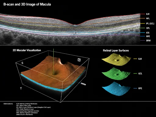
Spectral Domain-Optical Coherence Tomography (SD-OCT) Huvitz OCTavius

SD-OCT technology is used for fast and detailed retinal layer imaging

Fast

Non-invasive

High resolution 3D imaging

High sensitivity for detection of subtle changes in the retinal layers

Monitoring of disease progression and treatment response

Click on the image to zoom in

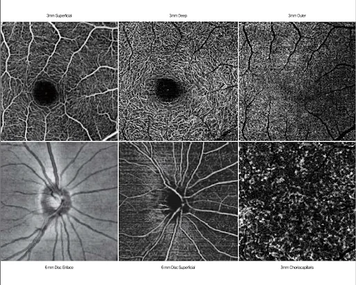
Optical Coherence Tomography- Angiography (OCTA) Huvitz OCTavius

OCTA technology allows for non- invasive (NO dye injection used) imaging of retinal and choroidal blood vessels

Fast

Non- invasive (No dye injection)

Early detection of abnormal vascular changes

Monitoring for disease progression and treatment response

Click on the image to zoom in

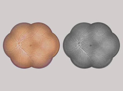
Fundus imaging

Colour retinal images optimised with high resolution and contrast are useful for clinical diagnosis

Better visualisation and documentation of abnormalities

Monitor the progression or regression of lesions on follow up

Click on the image to zoom in

Treatment modalities at Namah

01

Medical management

Medical management in uveitis is done in the form of eye drops or oral medication. Detailed systemic evaluation by special blood tests helps to determine the underlying cause and start targeted systemic therapy.

02

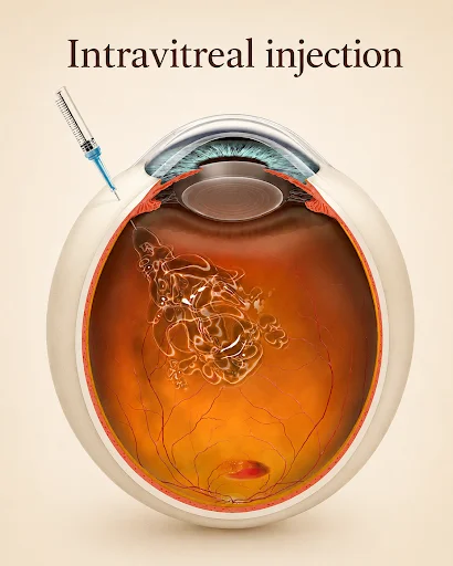
Intravitreal or periocular injections

It is a minimally invasive procedure to deliver potent medicines inside the eye for maximum benefit to the patient.

Procedure is quick, safe and is performed under topical anesthesia (using eye drops).

It is given for retinal swelling or due to presence of abnormal new blood vessels or infections. These conditions include macular edema, endophthalmitis, occlusive proliferative diseases or other inflammatory conditions.

At Namah, this procedure is performed in the modular operation theatre under aseptic precautions to ensure maximum safety for the patient.

Click on the image to zoom in

03

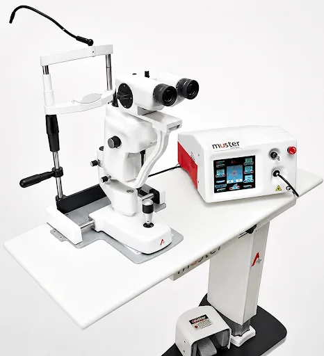
RETINAL LASER AT NAMAH

Non-invasive LASER retinal treatment is a fast and safe OPD procedure done under topical anesthesia (using eye drops)

Certain inflammatory conditions resulting in non-perfused retinal areas or proliferative changes require retinal LASER treatment

At Namah, multi-spot green laser (AEON meditech) is used to deliver multiple laser spots at one time, ensuring quick laser procedure completion with minimal pain for the patient.

Click on the image to zoom in

04

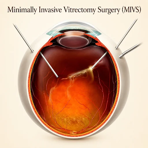
VITRECTOMY SURGERY AT NAMAH

Advanced Minimally Invasive Vitrectomy Surgery (MIVS) is performed in advanced cases of uveitis causing vitreo-retinal complications.

In some cases, silicone oil or gas is placed inside the eye during surgery to support the retina. Silicone oil removal may then be required after a few months.

Need for special post- operative care in the form of head positioning may be needed in certain cases.

Click on the image to zoom in

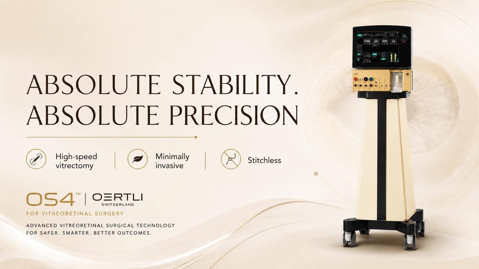
Oertli OS 4 vitrectomy platform

An advanced surgical platform is used for vitrectomy surgery at Namah

High speed vitrectomy with precise and controlled surgical environment

Highly stable ocular dynamics ensuring improved surgical safety

Minimally invasive procedure- MIVS incisions are very small and usually do not require any sutures

Faster healing

Better visual outcomes

Click on the image to zoom in

Frequently Asked Questions

Yes. Some types of uveitis can recur or become chronic. Follow-up is important even after symptoms improve.

Sometimes. Uveitis may be associated with infections, autoimmune conditions or inflammatory disease elsewhere in the body. Selected patients may need blood tests or physician/rheumatology coordination.

Scroll to top